Revista
Autotransplantation: A clinical option for ankylosed teeth
Miguel Roig Cayón
Doctor en Medicina, Estomatólogo. Jefe de Área de Restauración Dental y Endodoncia
José Espona Roig
Odontólogo. Profesor Asociado, Máster en Estética Dental. Profesor Asociado. Área de Restauración Dental y Endodoncia.
Joan Grau Cases
Cirujano máxilo-facial. Odontólogo. Profesor Asociado. Área de Cirugía Oral e Implantología
Fernando Durán-Sindreu Terol
Doctor en Odontología. Máster en Endodoncia. Director del Máster en Endodoncia. Área de Restauración Dental y Endodoncia
Ankylosis of anterior teeth is a serious complication of replanted or severely traumatized permanent incisors. These processes might cause a severe damage to the periodontal ligament cells that cover the root surface. As a result of this damage, the periodontal ligament is replaced by bone tissue, causing ankylosis between tooth and bone1. Once ankylosis is established it will eventually continue until complete resorption of the root and replacement by bone, leading to tooth loss. Ankylosis and replacement resorption are largely responsible of the low 5-year survival of teeth after these injuries2-4. According to Andersson5 the rate of root resorption in the group of patients aged 8 to16 years was significantly higher compared with the group of patients aged 17 to 39 years.
Most of dental trauma conducing to avulsion, intrusion and lateral luxation occur to young individuals, being most common between 8 and 10 year old patients, during the early mixed dentition6,7. In a growing individual ankylosis will develop an infraposition of the damaged tooth. The alveolar development will be arrested and the future aesthetic and/or functional treatment will be severely compromised. Therefore, an ankylosed tooth in those conditions should be removed before future orthodontic and/or prosthodontic therapy is jeopardized8. This should be done as soon as infraposition in diagnosed.
Treatment options to be considered include: composite builp-up, bone distraction, segmental osteotomy, tooth extraction, decoronation and auto transplantation. Tooth extraction seems to be an inadequate treatment option for the undesirable lowering and diminishing of the horizontal and vertical volume that it always causes9-11, in this case increased by the probably bone loss related to the always difficult extraction of an ankylosed tooth. And implant placement in the area has to be necessarily postponed until complete growth of the patient has been accomplished. Correcting the defect with a composite build up might be a treatment option if the patient has passed the pubertal growth and there is minimal infraposition, but it would never be recommended for growing patients, for infraposition will be increased, and the patient will end with a very long tooth and big asymmetry in the gingival margins. Bone distraction by dento-osseus osteotomy of the segment would be a possibility12-14 but it should be postponed until complete growth of the patient. Segmental osteotomy with autogenous bone graft has also been proposed, but there is only one case presented15, as far as we could find. Therefore, only two options remain, decoronation of the ankylosed tooth and auto transplantation of the first premolar to the anterior region.
Decoronation is a simple and safe surgical method first described by Malmberg in 198416 for preservation of alveolar bone prior to implant placement. As soon as infraocclusion is detected, a flap is raised to gain good access. The crown is removed two millimetres below the cervical margin. An endodontic file or a bur are then used to remove any root filling material or pulp from the root canal, and to allow blood flow into root canal space. The flap is then reattached covering the width of the tooth. When decoronation is done before pubertal growth spurt a vertical bone growth might be expected, and the width of the alveolar ridge is maintained in patients of all ages8. It is probably the treatment of choice in most of ankylosed teeth in growing patients for it preserves alveolar dimension thus enabling implantation after the completion of growth and development.
An alternative to decoronation is tooth auto transplantation. Many of the patients suffering ankylosis in anterior teeth have malocclusion17, mainly Angle class II, and might need tooth extractions to correct their malocclusion. If the orthodontic study determines the need of lower premolar extractions, auto transplantation should be considered. Auto transplantation is a reliable treatment method. In a study Kvint et al. informed of an overall success rate of 81% over 215 patients18.
The highest success rate, a 100%, was for transplantation of premolars to the maxillary anterior region. According to Czochrowska et al.19 the overall status of the transplanted premolars and surrounding tissues, similar to natural incisors, indicated that this treatment modality may be recommended when maxillary incisors are missing in adolescents. In addition, tooth transplantation represents an inherent potential for bone induction and reestablishment of a normal alveolar process.
Case description
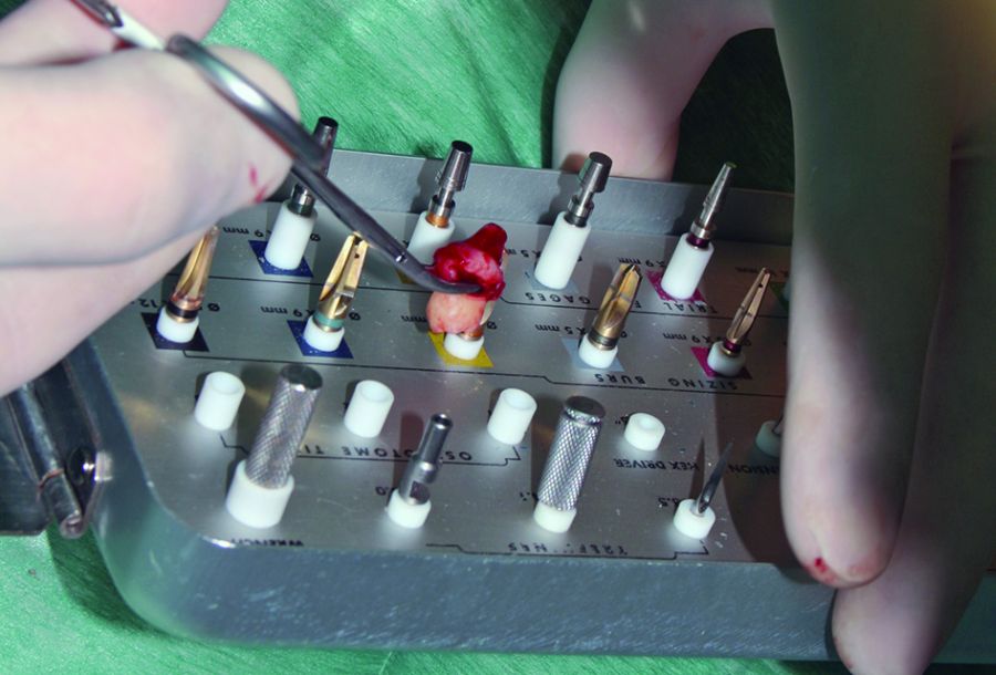 1. With the extracted tooth we select the best bur to prepare the socket. The extracted tooth will then be kept in cold saline until insertion in the prepared socket.
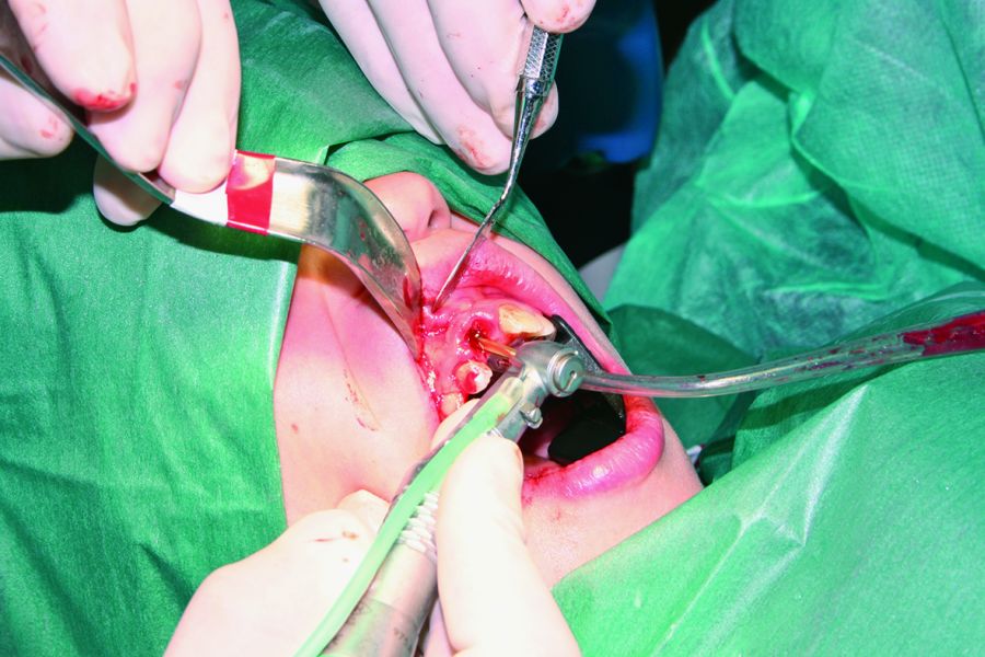 2. The socket is prepared by means of the selected implant bur at low speed.
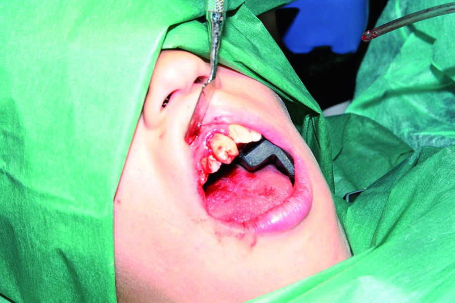 3. Inital positioning of the mandibular premolar in the incisor socket, to test adaptation.
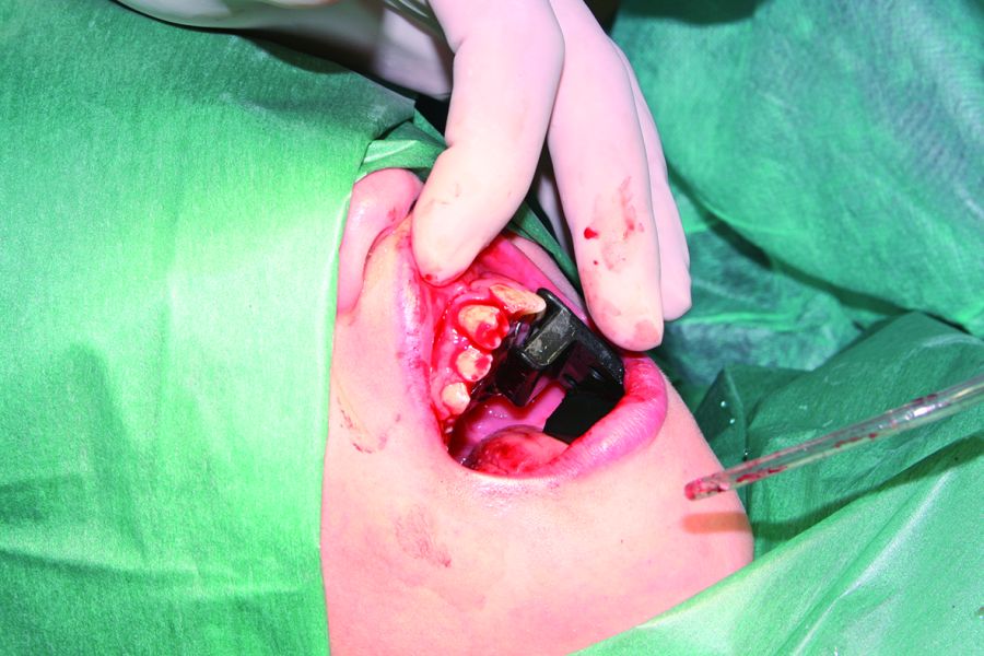 4. The auto transplanted tooth is left in slight infraocclusion.
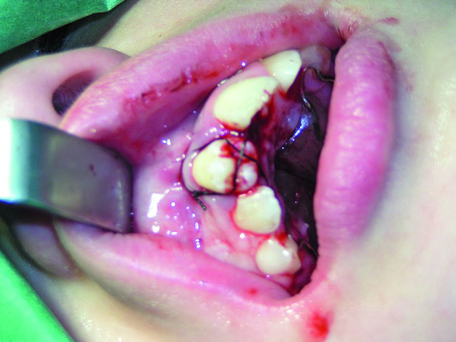 5. A suture was used to secure tooth position.
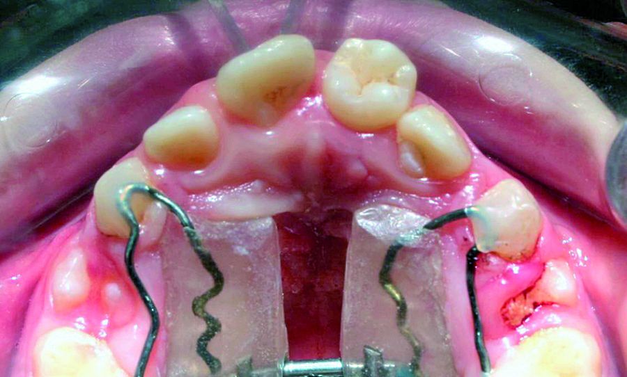 6. Occlusal view of autransplanted tooth one month after the surgical procedure.
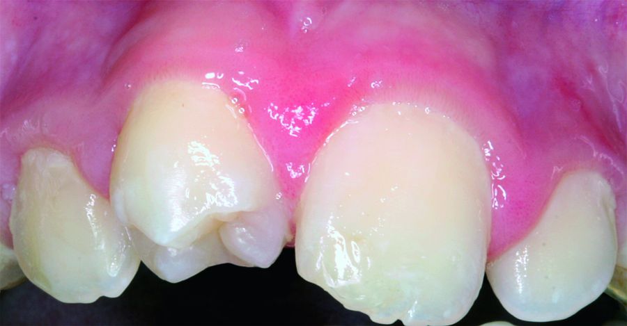 7. Tooth 1.1(4.4.) one month after tooth autotransplantation.
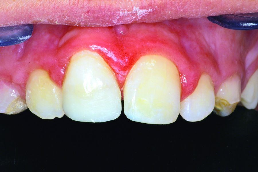 8. Two months after autotransplantation, tooth is prepared and composite is used to improve aesthetic appareance.
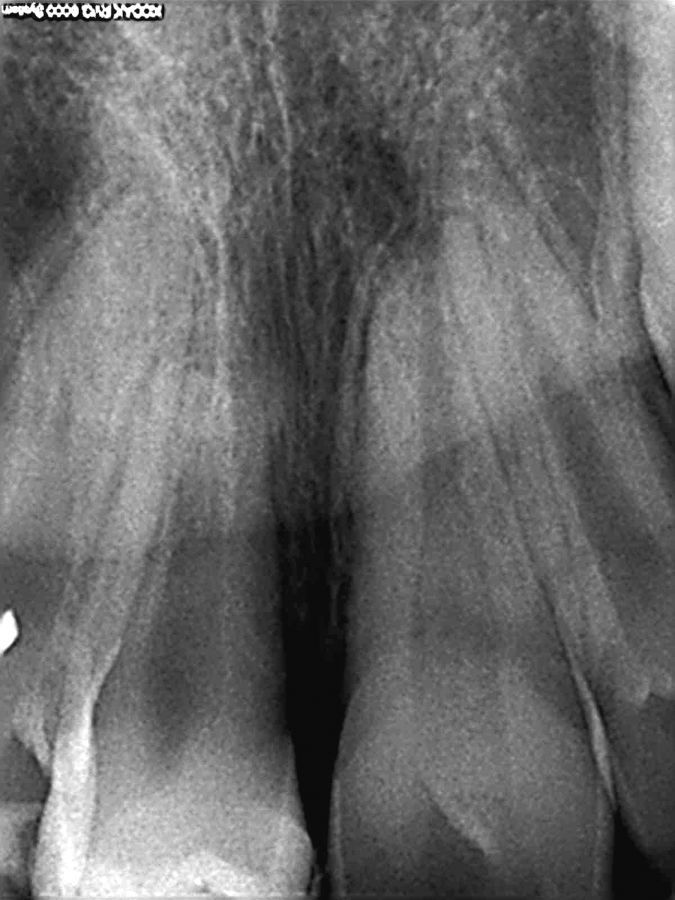 9.Tooth root two years after auto transplantation.
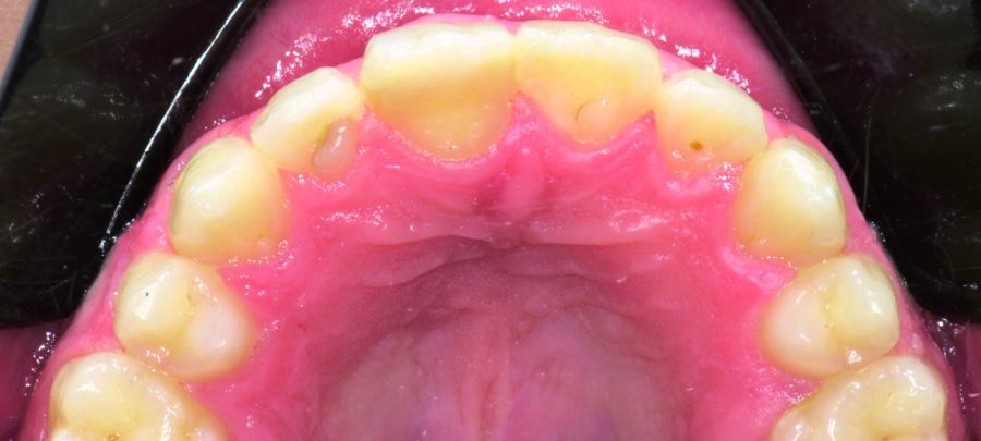 10. Occlusal view of autotransplanted tooth after 10 years. The patient is now 20 year old.
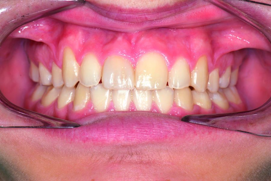 11. Buccal view after 10 years. The autotransplanted tooth is slighty narrower and the cenit is a little bit lower. In order to achive better aesthetic result a gingival recontouring and veneering might be recommended after complete growth of the patient.
A 10 years old patient was referred to Dental School clinic after avulsion of tooth 1.1. The tooth had been out of the mouth and dry stored for over two hours. Although the prognosis of the tooth was poor, replantation was done.
The alveolar socket was thoroughly rinsed with saline. The tooth root surface was also cleaned with saline, and immersed for 20 minutes in 2% sodium fluoride. The tooth was then replanted, and splinted with a 0.014 orthodontic wire to neighbour teeth. Amoxicillin plus clavulanic acid was prescribed for 7 days. Tetanus booster was applied20.
The patient was referred to the orthodontic department, to have an orthodontic study done. The need of orthodontic treatment with full braces and extraction of two maxillary and mandibular first premolars was established. As ankylosis was expected to happen, the decision of auto transplantation of an upper premolar in place of tooth 11 was taken. At one-year control infraposition of tooth 1.1 was clearly observed. If auto transplantation had not been decided, it would have been necessary to proceed with decoronation at that time. As auto transplantation was the treatment option of choice, the growth of the first lower premolars roots was controlled every six months until two thirds of the root of one of them was developed21. When that was observed, we proceeded with auto transplantation.
For auto transplantation, tooth 1.1 was carefully removed. The right first lower premolar was carefully extracted, raising a flap in order to better access the incompletely erupted tooth, and the tooth was kept in cold saline. From the different implant burs available in the clinic, the one that better fit with the root width was selected (Figure 1). The bone was drilled with the selected implant bur to create an artificial alveolar socket where to replant the premolar (Figure 2).
The extraoral time lapse for the autotransplanted tooth is critical for success. For that reason it has been recommended to produce a resin model of the tooth to be transplanted from a CT image, so that the adaptation of the resin model to the transplantation socket is finished before the tooth to be transplanted is extracted, minimizing in that way the extraoral time lapse22.
At the time of this treatment that option was not yet available. The root of the extracted premolar was measured to determine if it would perfectly fit in the prepared socket (Figure 3). As the measure of the socket was correct, the tooth was directly placed in the artificial alveolar socket. As the mesio-distal diameter was smaller than the bucco-lingual diameter, the premolar was rotated prior to replantation, to allow a more aesthetic reconstruction in the future.
The replanted premolar was left slightly in infraocclusion (Figure 4), and suture was placed to secure its position (Figure 5). Suture was removed after 7 days.
Two months after auto transplantation (Figures 6, 7), the palatal aspect of the tooth was prepared to give it a palatal anatomy similar to that of a normal central incisor. The exposed dentin was covered with composite in order to protect it. Composite was also used to give a buccal morphology similar to that of a central incisor (Figure 8).
In the follow up it was clearly seen how the transplanted tooth erupted until occlusion was achieved, and from them on the gingival margins were kept at the same level in respect to neibourghing teeth. The root length slightly increased in between the first two years (Figure 9).
Pulp space was obliterated, what seems to be a sign of revascularization. Two years after tooth auto transplantation, the patient was summited to orthodontic treatment with braces.
A three year treatment was completed, and many teeth, the auto transplanted tooth included, were moved, with neither signs nor symptoms of pathology.
At the 10-year control auto transplanted tooth remained stable, without signs or symptoms of any pathology (Figure 10). From an aesthetic point of view there might be a need of gingival recontouring and a cosmetic composite or laminate veneer (Figure 11). Nevertheless the patient refused any further treatment for the moment.
Correspondence:
mroig@uic.es
Bibliografía/References
1. Levin I, Ashkenazi M, Schwartz-Arad D. Preservation of alveolar bone of un-restorable traumatized maxillary incisors for future. Refuat Hapeh Vehashinayim 2004;21(1):54-59,101-102.
2. Barrett EJ, Kenny DJ. Survival of avulsed permanent maxillary incisors in children following delayed replantation. Endod Dent Traumatol 1997;13(6):269-275.
3. Nikoui M, Kenny DJ, Barrett EJ. Clinical outcomes for permanent incisor luxations in a pediatric population. III. Lateral luxations. Dent Traumatol 2003;19(5):280-285.
4. Humphrey JM, Kenny DJ, Barrett EJ. Clinical outcomes for permanent incisor luxations in a pediatric population. I. Intrusions. Dent Traumatol 2003;19(5):266-273.
5. Andersson L, Bodin I, Sorensen S. Progression of root resorption following replantation of human teeth after extended extraoral storage. Endod Dent Traumatol 1989;5(1):38-47.
6. Malmgren B. Decoronation: how, why, and when? J Calif Dent Assoc 2000;28(11):846-854.
7. Andreasen JO, Ravn JJ. Epidemiology of traumatic dental injuries to primary and permanent teeth in a Danish population sample. Int J Oral Surg 1972;1(5):235-239.
8. Malmgren B. Ridge preservation/decoronation. J Endod 2013;39(3 Suppl):S67-72.
9. Araujo MG, Lindhe J. Dimensional ridge alterations following tooth extraction. An experimental study in the dog. J Clin Periodontol 2005;32(2):212-218.
10. Schropp L, Wenzel A, Kostopoulos L, Karring T. Bone healing and soft tissue contour changes following single-tooth extraction: a clinical and radiographic 12-month prospective study. Int J Periodontics Restorative Dent 2003;23(4):313-323.
11. Pietrokovski J, Massler M. Alveolar ridge resorption following tooth extraction. J Prosthet Dent 1967;17(1):21-27.
12. Kofod T, Wurtz V, Melsen B. Treatment of an ankylosed central incisor by single tooth dento-osseous osteotomy and a simple distraction device. Am J Orthod Dentofacial Orthop 2005;127(1):72-80.
13. Isaacson RJ, Strauss RA, Bridges-Poquis A, Peluso AR, Lindauer SJ. Moving an ankylosed central incisor using orthodontics, surgery and distraction osteogenesis. Angle Orthod 2001;71(5):411- 418.
14. Medeiros PJ, Bezerra AR. Treatment of an ankylosed central incisor by single-tooth dentoosseous osteotomy. Am J Orthod Dentofacial Orthop 1997;112(5):496-501.
15. You KH, Min YS, Baik HS. Treatment of ankylosed maxillary central incisors by segmental osteotomy with autogenous bone graft. Am J Orthod Dentofacial Orthop 2012;141(4):495-503.
16. Malmgren B, Cvek M, Lundberg M, Frykholm A. Surgical treatment of ankylosed and infrapositioned reimplanted incisors in adolescents. Scand J Dent Res 1984;92(5):391-399.
17. Borzabadi-Farahani A. The association between orthodontic treatment need and maxillary incisor trauma, a retrospective clinical study. Oral Surg Oral Med Oral Pathol Oral Radiol Endod 2011;112(6):e75-80.
18. Kvint S, Lindsten R, Magnusson A, Nilsson P, Bjerklin K. Autotransplantation of teeth in 215 patients. A follow-up study. Angle Orthod 2010;80(3):446-451.
19. Czochrowska EM, Stenvik A, Album B, Zachrisson BU. Autotransplantation of premolars to replace maxillary incisors: a comparison with natural incisors. Am J Orthod Dentofacial Orthop 2000;118(6):592-600.
20. The dental trauma guide. 2010 ; Available from: http://www. dentaltraumaguide.org/Permanent_Avulsion_Treatment.aspx.
21. Mendoza-Mendoza A, Solano-Reina E, Iglesias-Linares A, Garcia-Godoy F, Abalos C. Retrospective long-term evaluation of autotransplantation of premolars to the central incisor region. Int Endod J 2012;45(1):88-97.
22. Lee SJ, Kim E. Minimizing the extra-oral time in autogeneous tooth transplantation: use of computer-aided rapid prototyping (CARP) as a duplicate model tooth. Restor Dent Endod 2012;37(3):136-141.

La Inteligencia Artificial implementada en sistemas que permiten el control de determinados tratamientos garantiza la continuidad de estos durante el verano, incluso a distancia.

La Asamblea Mundial de la Salud (WHA78) concluyó con una nota constructiva, con varios acuerdos mundiales notables para promover la equidad en la salud y la resiliencia de los sistemas de salud en todo el mundo.

La detección y corrección temprana de hábitos como la respiración bucal o la masticación inadecuada puede reducir significativamente la necesidad de ortodoncia en etapas posteriores del desarrollo infantil.

Pfizer España ha celebrado la III edición de “Esto es ciencia, no ficción”, una iniciativa orientada a divulgar conocimiento riguroso y acercar a la sociedad la ciencia más innovadora y sus últimos avances.

La compañía ha registrado un aumento interanual tanto en el número de cursos de formación clínica ofrecidos a nivel mundial como en el número de profesionales dentales que participaron en ellos.

Sus múltiples usos podrían contribuir a mejorar diagnósticos, personalizar tratamientos en base a las características de cada paciente y optimizar la gestión de recursos contribuyendo a una gestión más eficiente de los servicios de salud.

Un grupo de investigadores de la Universidad de Ciencia y Tecnología de Huazhong en Wuhan (China), ha realizado un análisis exhaustivo de las estrategias actuales y futuras para la regeneración del complejo pulpo-dentinario, con el objetivo de sustituir la terapia de conductos tradicional por soluciones biológicamente activas capaces de restaurar la vitalidad dental.

Bajo el lema "Abriendo puertas a la colaboración científica", la compañía celebró un encuentro centrado en difundir ciencia e investigación.

Donte Group se convierte en la primera empresa de salud de bucodental en recibir la Certificación Top Employers 2025.
